 More and more research has raised concern over the dangers of concussions – one of the most common forms of head trauma – as many sufferers go on to experience persistent neurological symptoms throughout their lives.
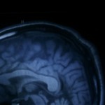
Now, scientists have discovered a clue as to why mild traumatic brain injuries (MTBI) can have such long-lasting health consequences.
In a study published in the journal Radiology, researchers found that white matter damage in the brains of people who had experienced concussions closely resembled the type of white matter damage found in patients with Alzheimer’s disease. These findings suggest that concussions set off a chain of neurological events that can cause long-term damage to the brain.
“It’s not the hitting your head that’s the problem. It’s everything else that happens after that,” said lead study author Dr. Saeed Fakhran, assistant professor of radiology in the Division of Neuroradiology at the University of Pittsburgh School of Medicine.
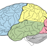 Primary types of neuromodulation techniques used in psychiatry and new approaches to treat neurological and psychiatric problem.
 New guidelines published online May 6 in the Canadian Medical Association Journal have codified a number of major changes in fibromyalgia (FM) diagnosis and treatment. The authors of the new guidelines note that FM diagnosis and care have largely shifted to primary care physicians and away from rheumatologists and other specialists.
 At a press conference held at the American Academy of Neurology’s (AAN’s) 2013 Annual Meeting, the release of new AAN guidelines for the evaluation and management of sports-related concussion (SRC) were announced. The recommendations update the 1997 AAN sports concussion practice parameter and were published online in Neurology on March 18, 2013.[1] The new guidelines attempt to address uncertainty and inconsistency in the management of concussion and mild traumatic brain injury (TBI) by addressing 4 clinical questions:
 Stroke is a common, potentially devastating disease with potential high morbidity and mortality. Recognition at the onset of acute ischemic stroke is pivotal to changing outcomes such as intravenous thrombolysis. Stroke monitoring is a burgeoning field with various methods described and newer devices that aid in detecting acute or worsening ischemia that can lead to improved bedside and intensive care unit management. This article describes various methods of bedside stroke monitoring including newer techniques of intracranial pressure monitoring using the pressure reactivity index and compensatory reserve index to detect changes in autoregulatory states, noninvasive intracranial pressure monitoring, quantitative EEG with alpha–delta ratio, transcranial Doppler, methods of arteriovenous brain oxygen monitoring such as jugular venous oxygen and near-infrared spectroscopy, invasive brain oxygen probes such as LicoxTM (brain tissue O2), cerebral blood flow probe (CBF HemedexTM) and cerebral microdialysis.